45 cell membrane diagram with labels
PDF Human Cell Diagram, Parts, Pictures, Structure and Functions The cell membraneis the outer coating of the cell and contains the cytoplasm, substances within it and the organelle. It is a double-layered membrane composed of proteins and lipids. The lipid molecules on the outer and inner part (lipid bilayer) allow it to selectively transport substances in and out of the cell. Endoplasmic Reticulum CELL MEMBRANE LABEL Diagram | Quizlet Practice labeling the parts of the cell membrane Terms in this set (6) Channel Protein hole or tunnel that particles may pass through to go in / out of cell Marker protein identifies or labels the cell Receptor protein receives information Heads part of the phospholipid that loves water (hydrophili) - points to the most outside and inside of cell
Plasma Membrane Function, Structure & Diagram - Study.com 2. property of the plasma membrane that allows some substances into the cell and keeps others out 4. main structural component of the plasma membrane 6. nonpolar part of a phospholipid 11. protein...

Cell membrane diagram with labels
Labeling Cell Membrane - Labelled diagram - Wordwall Labeling Cell Membrane - Labelled diagram. integral protein, cholesterol, external cell environment, hydrophilic part of phospholipid bilayer, peripheral protein, internal environment of the cell, hydrophobic part of phospholipid bilayer, glycolipid. Labeled Plant Cell With Diagrams | Science Trends The parts of a plant cell include the cell wall, the cell membrane, the cytoskeleton or cytoplasm, the nucleus, the Golgi body, the mitochondria, the peroxisome's, the vacuoles, ribosomes, and the endoplasmic reticulum. Parts Of A Plant Cell The Cell Wall Let's start from the outside and work our way inwards. PDF Label The Cell Membrane Diagram Ivyspring Com Terms For Full Terms And Conditions''cell Membrane Coloring Home Biology Junction May 6th, 2018 - NAME DATE PERIOD the cell membrane structure or its function with the correct letter from the cell membrane diagram Color and label the cell in an' 'Interactive Cell Models May 5th, 2018 - This introduction to the structure of plant ...
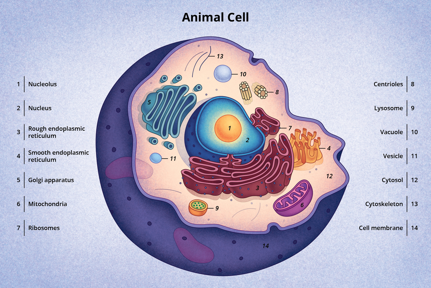
Cell membrane diagram with labels. Cell Membrane Function and Structure - ThoughtCo The cell membrane is a multifaceted membrane that envelopes a cell's cytoplasm. It protects the integrity of the cell along with supporting the cell and helping to maintain the cell's shape. Proteins and lipids are the major components of the cell membrane. The exact mix or ratio of proteins and lipids can vary depending on the function of a ... Cell Membrane 3 D Model With Label Labeled : Functions and Diagram Regarding the term 'fluid mosaic model', the cell membrane is more like a fluid, rather than being a rigid or solid structure. You can use ordinary household objects to represent these organelles, it doesn't have to be expensive. Things to include & label in your drawing: Phospholipids o Hydrophilic heads o Hydrophobic tails • Cytoplasm. Cell Membrane Structure Labeled Labeled : Functions and Diagram Cell Membrane Structure Labeled. Let us study the detailed composition of this lipid bilayer and other substances found in the cell membrane. This structure has two layers, and is represented in the diagram below. We all do not forget that the human physique is very intricate and one way I learned to comprehend it is by way of the style of human ... Animal Cell Diagram with Label and Explanation: Cell ... - Collegedunia Animal cells are eukaryotic cells. They contain membrane-bound nuclei. Diagram of Animal Cell is beneficial in understanding the structure and functions of an animal. This article comprehends a brief explanation of the different parts of an animal cell with a well-labelled diagram. Table of Content.
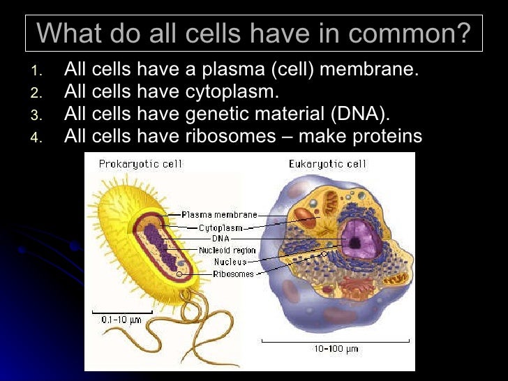
Diagram of a cell membrane with labels | NIST Biology in Reflectometry. Essential Biological Functions. Immune response, Cell metabolism, Neurotransmission, Photosynthesis, Cell adherence, Cell growth and differentiation. Potential Commercial Applications. Drug response monitoring, Chemical manufacturing, Biosensing, Energy conversion, Tissue engineering. Animal Cells: Labelled Diagram, Definitions, and Structure Plant cells have chloroplasts to synthesize their own food. Absent: Plasma Membrane: Cell wall and a cell membrane: Only cell membrane: Flagella: Present in some cells (e.g. sperm of bryophytes and pteridophytes, cycads and Ginkgo) Present in some cells ( e.g. mammalian sperm cells) Cilia: Most plant cells do not contain cilia. Present: Lysosomes Cell: Structure and Functions (With Diagram) - Biology Discussion Eukaryotic Cells: 1. Eukaryotes are sophisticated cells with a well defined nucleus and cell organelles. 2. The cells are comparatively larger in size (10-100 μm). 3. Unicellular to multicellular in nature and evolved ~1 billion years ago. 4. The cell membrane is semipermeable and flexible. 5. These cells reproduce both asexually and sexually. en.wikipedia.org › wiki › Electron_transferElectron transfer - Wikipedia Electron transfer (ET) occurs when an electron relocates from an atom or molecule to another such chemical entity. ET is a mechanistic description of certain kinds of redox reactions involving transfer of electrons.
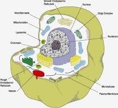
Human Cell Diagram, Parts, Pictures, Structure and Functions Diagram of the human cell illustrating the different parts of the cell. Cell Membrane. The cell membrane is the outer coating of the cell and contains the cytoplasm, substances within it and the organelle. It is a double-layered membrane composed of proteins and lipids. The lipid molecules on the outer and inner part (lipid bilayer) allow it to ... File:Cell membrane detailed diagram 4.svg - Wikipedia Cell membrane detailed diagram 4.svg. English: The cell membrane, also called the plasma membrane or plasmalemma, is a semipermeable lipid bilayer common to all living cells. It contains a variety of biological molecules, primarily proteins and lipids, which are involved in a vast array of cellular processes. quizlet.com › 244659812 › cell-bio-ch-22-flash-cardsCell Bio - Ch. 22 Flashcards | Quizlet The diagram below shows the five main transport proteins that control the distribution of Na+ and K+ ions across the plasma membrane of an axon. Assume that the membrane is at resting potential---the membrane potential of the axon remains constant at about -70 mV. › cell_cycle_jsInteractive Cell Cycle - CELLS alive INTERPHASE. Gap 0. Gap 1. S Phase. Gap 2. MITOSIS . ^ Cell Cycle Overview Cell Cycle Mitosis > Meiosis > Get the Cell Division PowerPoints
Explain the nucleus of a cell with a neat labeled diagram - Science - Cell - Structure and ...
Cell Membrane Diagram Teaching Resources | Teachers Pay Teachers This diagram resource makes learning about parts of a cell membrane easy and engaging! A detailed handout with labels is provided, as well as a fillable worksheet.The handout and worksheet is provided in both color and black and white.This resource can be printed, or used digitally via the included Easel activity!
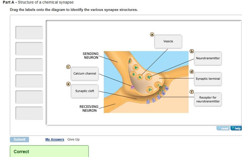
Chapter 11 - Neurophysiology Activities Flashcards | Easy Notecards
File:Cell membrane detailed diagram blank.svg - Wikimedia Description. Cell membrane detailed diagram blank.svg. English: The cell membrane, also called the plasma membrane or plasmalemma, is a semipermeable lipid bilayer common to all living cells. It contains a variety of biological molecules, primarily proteins and lipids, which are involved in a vast array of cellular processes.

Cell Membrane With Labels Functions - Cell Diagram
Labeling of Cell Membranes and Compartments for Live Cell Fluorescence ... The plasma membrane is the easiest cell membrane to label because it is very accessible. Fluorescent dyes and most commonly used dyes FM 1-43 family are used for labeling the plasma membrane. The cytosol is the intracellular space excluding the nucleus and the organelles.

Biology Concepts: Organelles
› books › NBK26894The Mitochondrion - Molecular Biology of the Cell - NCBI ... The enzymes of the respiratory chain are embedded in the inner mitochondrial membrane, and they are essential to the process of oxidative phosphorylation, which generates most of the animal cell's ATP. The inner membrane is usually highly convoluted, forming a series of infoldings, known as cristae, that project into the matrix. These ...

Organelles
Bacteria in Microbiology - shapes, structure and diagram Bacterial spores. Bacterial endospores layers. Bacteria cells are the smallest living cells that are known; even though viruses are smaller than bacteria, viruses are not living cells. There are different types of bacteria with various sizes, shapes, and structures. The bacteria shapes, structure, and labeled diagrams are discussed below.
Chapter 2 Cells Flashcards | Easy Notecards
Structure of Bacterial Cell (With Diagram) - Biology Discussion Cell wall: It is a tough and rigid structure of peptidoglycan with accessory specific materials (e.g. LPS, teichoic acid etc.) surrounding the bacterium like a shell and lies external to the cytoplasmic membrane. It is 10-25 nm in thickness. It gives shape to the cell. Nucleus: The single circular double-stranded chromosome is the bacterial genome.

Introduction to botany The Plant Cell
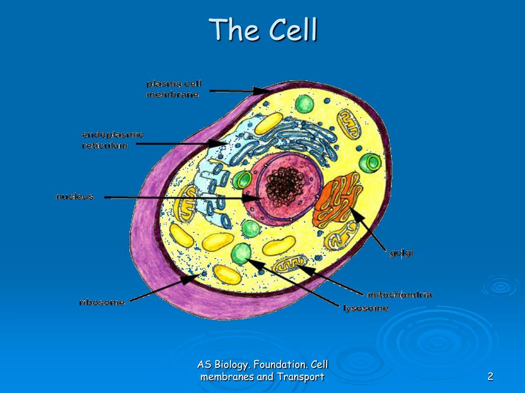
A Well-labelled Diagram Of Animal Cell With Explanation Well-Labelled Diagram of Animal Cell The Cell Organelles are membrane-bound, present within the cells. There are various organelles present within the cell and are classified into three categories based on the presence or absence of membrane. Listed below are the Cell Organelles of an animal cell along with their functions.
Cell Membrane Diagram Labeled : Functions and Diagram
A Labelled Diagram Of Mitochondria with Detailed Explanation Mitochondria are a double-membrane-bound cell organelle found in most eukaryotic organisms. In all living cells, these cell organelles are found freely floating within the cytoplasm of the cell. The diagram of Mitochondria is useful for both Class 10 and 12. It is one among the few topics having the highest weightage of marks and is majorly ...

Third Grade Science: Chapter 1 Plant and Animal Cells and Functions
Sell membrane | 140264jd | Flickr Some rights reserved. About; Jobs; Blog; Developers; Guidelines; Privacy; Terms; Help; SmugMug+Flickr.

Preface | Microbiology
Labeling Cell Membrane - Labelled diagram - Wordwall Labeling Cell Membrane - Labelled diagram receptor protein, phospholipid bilayer, external cell environment, phosphate group, Transport (channel protein), marker protein, internal environment of the cell, nonpolar part of the membrane. Labeling Cell Membrane Share by Walukas Like Edit Content More Leaderboard Log in required Theme Log in required

Cell Membrane Easy Definition Simple - Cell Diagram
Labeling a cell membrane Diagram | Quizlet Start studying Labeling a cell membrane. Learn vocabulary, terms, and more with flashcards, games, and other study tools.

CIE Biology Paper-4 Specimen Questions with Answers 1 to 2 - ExamTestPrep
biology4alevel.blogspot.com › 2015/08/95-using-re#96 Using respirometers | Biology Notes for A level - Blogger Aug 27, 2015 · Two rubber bungs are now taken, fitted with tubes as shown in the diagram. Close the spring clips. Attach the manometers to the bent glasstubing, ensuring an airtight connection. Next, place the bungs into the tops of the tubes. Open the spring clips. (This allows the pressure throughout the apparatus to equilibrate with atmospheric pressure.)
Cell Membrane What Does It Do Labeled - Cell Diagram
› articles › s41576/020/00292-xDeciphering cell–cell interactions and communication from ... Nov 09, 2020 · Cell–cell interactions orchestrate organismal development, homeostasis and single-cell functions. When cells do not properly interact or improperly decode molecular messages, disease ensues.
Discovery and Structure of Cells | Biology | Visionlearning
› cells › bactcellInteractive Bacteria Cell Model - CELLS alive They have an outer cell wall that gives them shape. Just under the rigid cell wall is the more fluid cell membrane. The cytoplasm enclosed within the cell membrane does not exhibit much structure when viewed by electron microscopy. Use the following animation to explore bacterial structure.

Cell Membrane Facts Labeled - Cell Diagram
Cell Organelles- Definition, Structure, Functions, Diagram A cell wall is multilayered with a middle lamina, a primary cell wall, and a secondary cell wall. The middle lamina contains polysaccharides that provide adhesion and allow binding of the cells to one another. After the middle lamina is the primary cell wall which is composed of cellulose.
Micro-organisms: Cells Flashcards | Easy Notecards
A Labeled Diagram of the Plant Cell and Functions of its Organelles The cell membrane is a thin layer made up of proteins, lipids, and fats. It forms a protective wall around the organelles contained within the cell. It is selectively permeable and thus, regulates the transportation of materials needed for the survival of the organelles of the cell. Function: Protects the cell from its surroundings. Cell Wall

Cell Analogy: A cell is like a hospital
Plant Cells: Labelled Diagram, Definitions, and Structure Plants have a rigid cell wall that surrounds the plasma membrane. The cell wall is made of cellulose and lignin, which are strong and tough compounds. Plant Cells Labelled Plastids and Chloroplasts Plants make their own food through photosynthesis. Plant cells have plastids, which animal cells don't.

BIOLOGY BLOG
PDF Label The Cell Membrane Diagram Ivyspring Com Terms For Full Terms And Conditions''cell Membrane Coloring Home Biology Junction May 6th, 2018 - NAME DATE PERIOD the cell membrane structure or its function with the correct letter from the cell membrane diagram Color and label the cell in an' 'Interactive Cell Models May 5th, 2018 - This introduction to the structure of plant ...
Post a Comment for "45 cell membrane diagram with labels"