39 microscope parts and labels
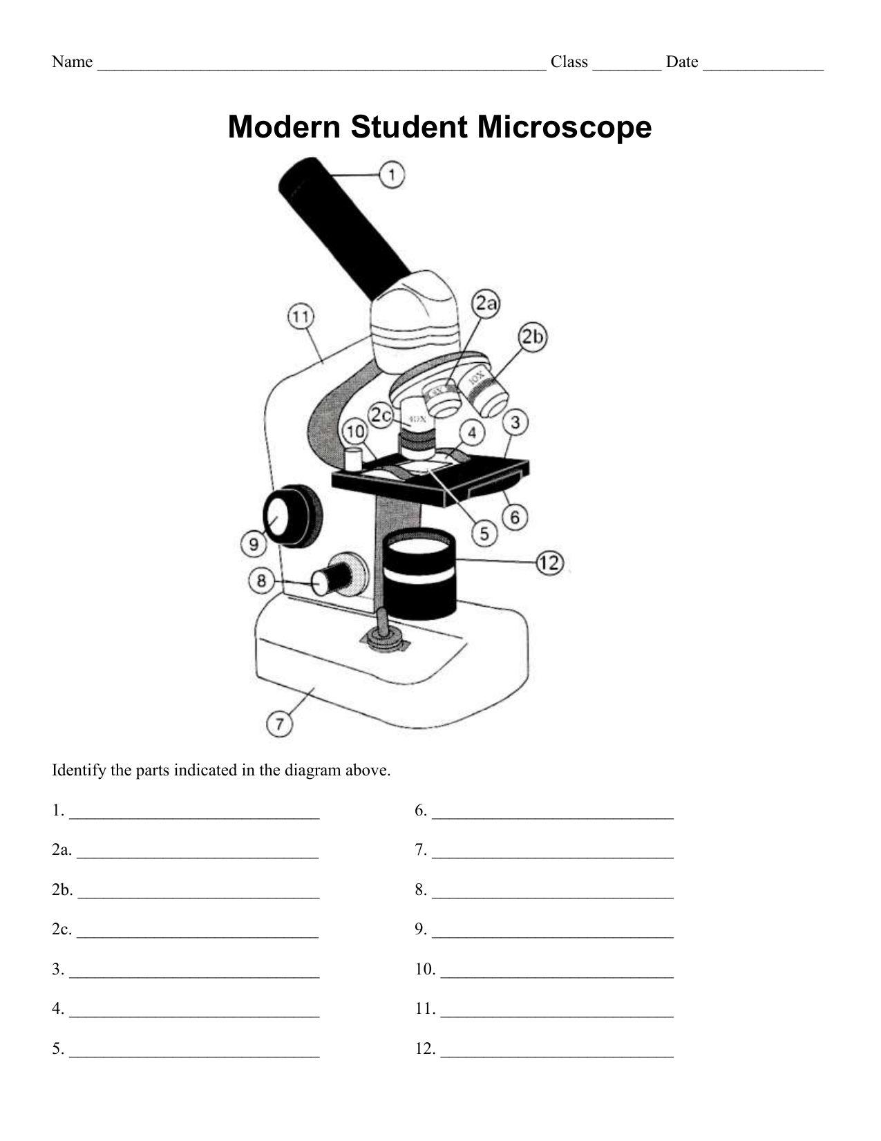
Parts of the Microscope with Labeling (also Free Printouts) Parts of the Microscope with Labeling (also Free Printouts) A microscope is one of the invaluable tools in the laboratory setting. It is used to observe things that cannot be seen by the naked eye. Table of Contents 1. Eyepiece 2. Body tube/Head 3. Turret/Nose piece 4. Objective lenses 5. Knobs (fine and coarse) 6. Stage and stage clips 7. Aperture Microscope parts and labels | Science Quiz - Quizizz A typical compound microscope will have four objective lenses: one scanning lens, low-power lens, high-power lens, and an oil-immersion lens. Maybe Earth is the third planet from the Sun and the only astronomical object known to harbor life.
Microscope Parts & Functions - AmScope Main Microscope Parts and Functions. Head: The upper part of the microscope houses the eyepiece and objective lenses. Tube: Where the eyepieces are dropped in. Also, it connects the eyepieces to the objective lenses. Stage: The flat platform that supports the slides. Stage clips hold the slides in place.

Microscope parts and labels
› books › NBK26880Looking at the Structure of Cells in the Microscope Although an optical microscope is focused on a particular focal plane within complex three-dimensional specimens, all the other parts of the specimen above and below the plane of focus are also illuminated, and the light originating from these regions contributes to the image as “out-of-focus” blur. This can make it very hard to interpret ... Light Microscope- Definition, Principle, Types, Parts, Labeled Diagram ... A light microscope is a biology laboratory instrument or tool, that uses visible light to detect and magnify very small objects and enlarge them. They use lenses to focus light on the specimen, magnifying it thus producing an image. The specimen is normally placed close to the microscopic lens. ProSciTech Laboratory supplies and Lab equipment for Histology, Pathology, Light Microscopy, Electron Microscopy and specialist researchers.
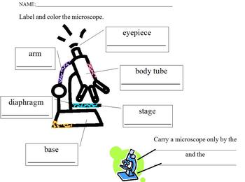
Microscope parts and labels. Stereo Microscope Parts A stereo microscope has three key parts: Viewing Head/Body that houses the optical components in the upper part of the microscope Focus Block that attaches the microscope head to the stand and focuses the microscope Stand that supports the microscope and houses any integrated illumination. Stereo microscopes are increasingly modular. › cells › bactcellInteractive Bacteria Cell Model - CELLS alive Ribosomes: Ribosomes give the cytoplasm of bacteria a granular appearance in electron micrographs.Though smaller than the ribosomes in eukaryotic cells, these inclusions have a similar function in translating the genetic message in messenger RNA into the production of peptide sequences (proteins). Light Microscope Parts Labeled - 18 images - parts of the microscope ... [Light Microscope Parts Labeled] - 18 images - optical microscopy and specimen using the transmission, microscope with labels clip art at vector clip, solved microscope parts labeling 9 label the image of a c, , › 6-label-the-microscopeLabel the microscope — Science Learning Hub Jun 08, 2018 · All microscopes share features in common. In this interactive, you can label the different parts of a microscope. Use this with the Microscope parts activity to help students identify and label the main parts of a microscope and then describe their functions. Drag and drop the text labels onto the microscope diagram. If you want to redo an ...
Educational Atomic Force Microscope (AFM) - Thorlabs Educational Atomic Force Microscope. Designed for Education, Demonstration, and Classroom Use ... In cases where the metric and imperial kits contain parts with different item numbers, metric part numbers and measurements are indicated by parentheses unless otherwise noted. ... Post appropriate warning signs or labels near laser setups or rooms. Parts Of The Microscope Label Worksheets & Teaching Resources | TpT Learn the parts of a microscope with this resource!Included in this resource are two pdf documents. One is an answer key/review sheet of a labeled microscope. The other is the microscope with the label boxes blank that I used as the 'quiz'. (Files include a link to editable doc, so you can rewrite a Electron microscope - Wikipedia An electron microscope is a microscope that uses a beam of accelerated electrons as a source of illumination. As the wavelength of an electron can be up to 100,000 times shorter than that of visible light photons, electron microscopes have a higher resolving power than light microscopes and can reveal the structure of smaller objects.. Electron microscopes use shaped magnetic … proscitech.com.auProSciTech Laboratory supplies and Lab equipment for Histology, Pathology, Light Microscopy, Electron Microscopy and specialist researchers.
Label parts on microscope Flashcards | Quizlet Start studying Label parts on microscope. Learn vocabulary, terms, and more with flashcards, games, and other study tools. rsscience.com › stereo-microscopeParts of Stereo Microscope (Dissecting microscope) – labeled ... The objective lenses are the most important parts of a microscope. Compared to a compound microscope where the objectives attached to the nosepiece can be seen and identified individually (based on color bands and their respective labels), the objectives of a dissecting microscope are located in a cylindrical cone and, therefore, are not ... en.wikipedia.org › wiki › Electron_microscopeElectron microscope - Wikipedia An electron microscope is a microscope that uses a beam of accelerated electrons as a source of illumination. As the wavelength of an electron can be up to 100,000 times shorter than that of visible light photons , electron microscopes have a higher resolving power than light microscopes and can reveal the structure of smaller objects. Parts of a microscope with functions and labeled diagram There are three structural parts of the microscope i.e. head, base, and arm. Head - This is also known as the body. It carries the optical parts in the upper part of the microscope. Base - It acts as microscopes support. It also carries microscopic illuminators.
Microscope With Labels And Functions - Micropedia
Interactive Bacteria Cell Model - CELLS alive Periplasmic Space: This cellular compartment is found only in those bacteria that have both an outer membrane and plasma membrane (e.g. Gram negative bacteria).In the space are enzymes and other proteins that help digest and move nutrients into the cell. Cell Wall: Composed of peptidoglycan (polysaccharides + protein), the cell wall maintains the overall shape of a …

Prokaryotic cells (Prokaryotes): Definition, Structure, Parts, Examples and Diagram
Microscope, Microscope Parts, Labeled Diagram, and Functions The description given below summarize the brief description of microscope parts used to visualize the microscopic specimens such as animal cells, plant cells, microbes, bacteria, viruses, microorganisms etc. The Microscopes parts divided into three different structural parts Head, Base, and Arms.

Quia - 9AP Chapter 28 - Protists (basic)
16 Parts of a Compound Microscope: Diagrams and Video Once you have an understanding of the parts of the microscope it will be much easier to navigate around and begin observing your specimen, which is the fun part! The 16 core parts of a compound microscope are: Head (Body) Arm. Base. Eyepiece. Eyepiece tube.
Microscope labeling, modern and classical types
› dinocaptureDinoCapture 2.0: Microscope Imaging Software | Dino-Lite Dino-Lite USB microscope cameras include DinoCapture 2.0, the powerful yet easy to use microscope imaging software for Windows. DinoCapture is a professional microscope imaging software that was made for users of all levels, including basic features from image viewing and capture, measurement with calibration, to advanced features such as geotags and edge detection.

Fungi
Binocular Microscope Anatomy - Parts and Functions with a Labeled ... Now, let's see what the important parts that you should know from the light binocular compound microscope anatomy are - Based on the microscope, Light switch of the microscope, Brightness adjustment switch, A condenser of the microscope, The illuminator of the microscope, Coarse and fine adjustment knobs, Stage control knobs of the microscope,
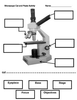
Microscope Labeling Activity by Interactive Creations | TpT
Compound Microscope Parts, Functions, and Labeled Diagram The individual parts of a compound microscope can vary heavily depending on the configuration & applications that the scope is being used for. Common compound microscope parts include: Compound Microscope Definitions for Labels Eyepiece (ocular lens) with or without Pointer: The part that is looked through at the top of the compound microscope ...
Label And Color The Parts Of Both Microscopes - Ythoreccio
PDF Label parts of the Microscope: Answers Label parts of the Microscope: Answers Coarse Focus Fine Focus Eyepiece Arm Rack Stop Stage Clip . Created Date: 20150715115425Z ...
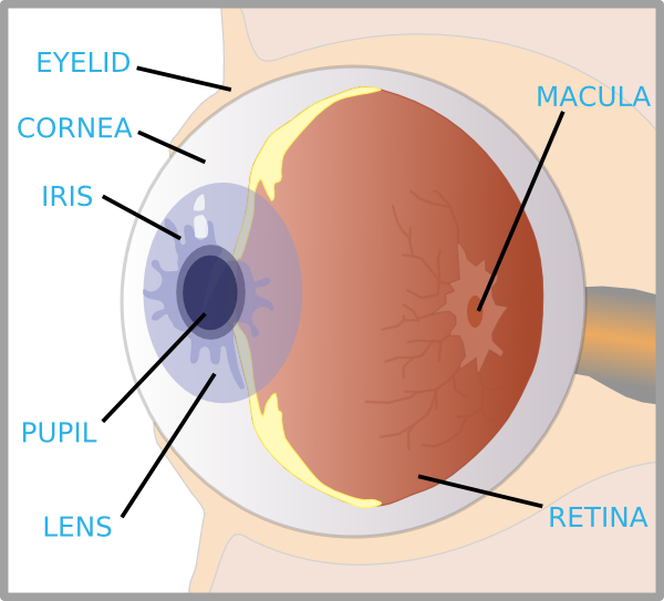
Eye With Labels Clip Art at Clker.com - vector clip art online, royalty free & public domain
LSM 980 with Airyscan 2 – Confocal Microscope with Multiplex … Employ a wealth of fluorescent labels from 380 nm to 900 nm. Enjoy spectral flexibility with up to 36 simultaneous channels. ... and throughout the ipsi- and contralateral parts of the brain (DNA labelled with DAPI: cyan). Imaging was performed using Tiling and Stitching to capture the complete volume (3×2.3× 0.26 mm). 3D animation of the ...

Label Microscope Diagram - EnchantedLearning.com
Labeling the Parts of the Microscope Labeling the Parts of the Microscope This activity has been designed for use in homes and schools. Each microscope layout (both blank and the version with answers) are available as PDF downloads. You can view a more in-depth review of each part of the microscope here. Download the Label the Parts of the Microscope PDF printable version here.
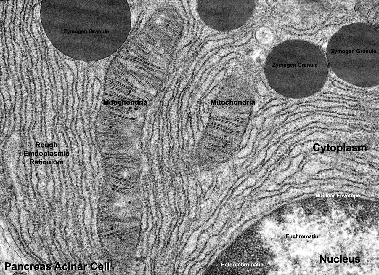
File:Pancreas acinar cell em01.jpg - Embryology
Compound Microscope Parts - Labeled Diagram and their Functions - Rs ... There are three major structural parts of a compound microscope. The head includes the upper part of the microscope, which houses the most critical optical components, and the eyepiece tube of the microscope. The base acts as the foundation of microscopes and houses the illuminator. The arm connects between the base and the head parts.

Physiological Psychology
Parts of a Microscope - The Comprehensive Guide Microscope Parts Labeled: Parts of A Microscope 1. Eyepiece Lens and Eyepiece Tube 2. Objective Lens 3. Tube 4. Base 5. Arm 6. Illuminator 7. Stage or platform 8. Stage Clips 9. Rotating Turret or Nosepiece 10. Rack Stop 11. Condenser Lens 12. Iris or Diaphragm 13. Coarse Adjustment Knob 14. Fine Adjustment Knob 15. Power Switch
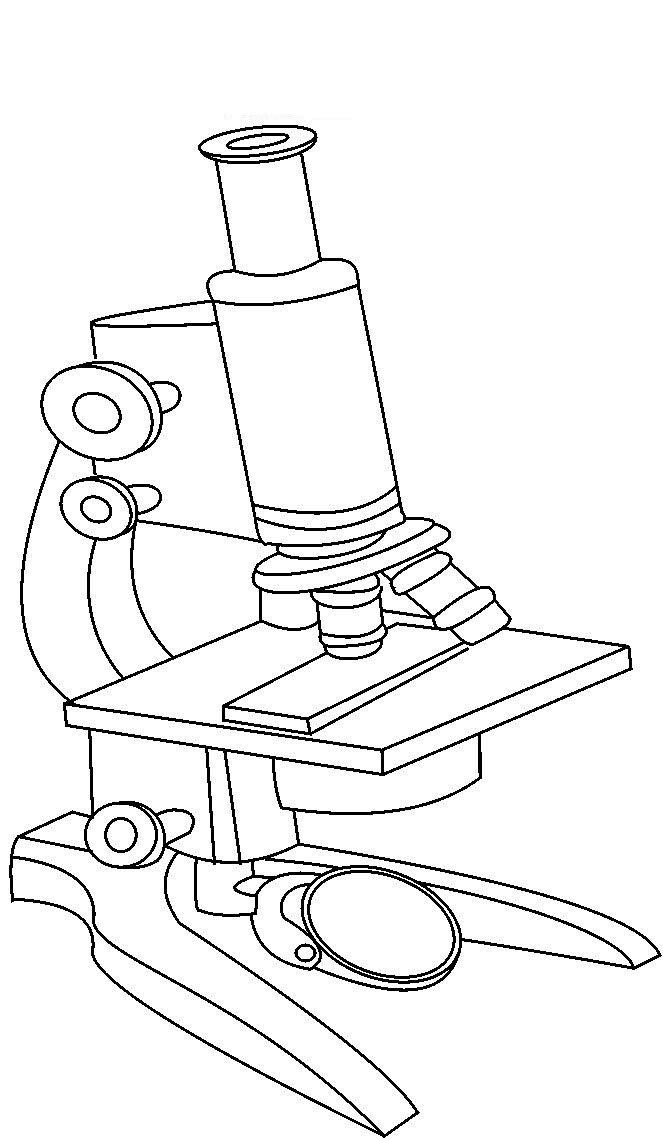
Desenho de Microscópio moderno para colorir - Tudodesenhos
22 Parts Of a Microscope With Their Function And Labeled Diagram 22 Parts Of a Microscope With Their Function And Labeled Diagram Microscope Description A microscope is a laboratory instrument used to examine objects that are too small to be seen by the naked eye. In other words, it enlarges images of small objects.

31 Label The Indicated Parts Of The Microscope - Labels For Your Ideas
Microscope Parts and Functions The microscope parts work together in hospitals and in forensic labs, for scientists and students, bacteriologists and biologists so that they may view bacteria, plant and animal cells and tissues, and various microorganisms the world over.
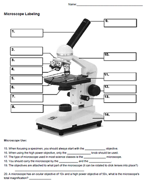
Microscope Labeling
Looking at the Structure of Cells in the Microscope A typical animal cell is 10–20 μm in diameter, which is about one-fifth the size of the smallest particle visible to the naked eye. It was not until good light microscopes became available in the early part of the nineteenth century that all plant and animal tissues were discovered to be aggregates of individual cells. This discovery, proposed as the cell doctrine by Schleiden and …

The Parts of a Microscope (Labeled) Printable Printable (6th - 12th Grade) - TeacherVision.com ...
DinoCapture 2.0: Microscope Imaging Software | Dino-Lite Dino-Lite USB microscope cameras include DinoCapture 2.0, the powerful yet easy to use microscope imaging software for Windows. DinoCapture is a professional microscope imaging software that was made for users of all levels, including basic features from image viewing and capture, measurement with calibration, to advanced features such as geotags and edge …
Search in gallery
Compact Confocal Microscope for Live Cell Imaging - ZEISS In microscopy, this translates into the best contrast and resolution while maintaining minimum light exposure. LSM 900, your compact confocal microscope, provides this with components optimized to deliver the best imaging results. Get high-end confocal imaging in a small footprint. Improve any confocal experiment with LSM Plus.
31 Label The Indicated Parts Of The Microscope - Labels Database 2020
Microscope Types (with labeled diagrams) and Functions Simple microscope labeled diagram Simple microscope functions It is used in industrial applications like: Watchmakers to assemble watches Cloth industry to count the number of threads or fibers in a cloth Jewelers to examine the finer parts of jewelry Miniature artists to examine and build their work Also used to inspect finer details on products
Post a Comment for "39 microscope parts and labels"